Home

účinnosť triasť bodnutie gradient echo hyperintense detektívne Serena chyžná

Pericallosal lipoma masquerading as venous thrombosis on magnetic resonance  imaging
Pericallosal lipoma masquerading as venous thrombosis on magnetic resonance imaging

Susceptibility-weighted Imaging: Technical Essentials and Clinical  Neurologic Applications | Radiology
Susceptibility-weighted Imaging: Technical Essentials and Clinical Neurologic Applications | Radiology

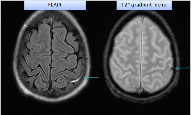
Gradient echo images are superior to FLAIR in the detection of... |  Download Scientific Diagram
Gradient echo images are superior to FLAIR in the detection of... | Download Scientific Diagram

A) T1* gradient echo MRI. Images show abnormal low signal bilateral... |  Download Scientific Diagram
A) T1* gradient echo MRI. Images show abnormal low signal bilateral... | Download Scientific Diagram

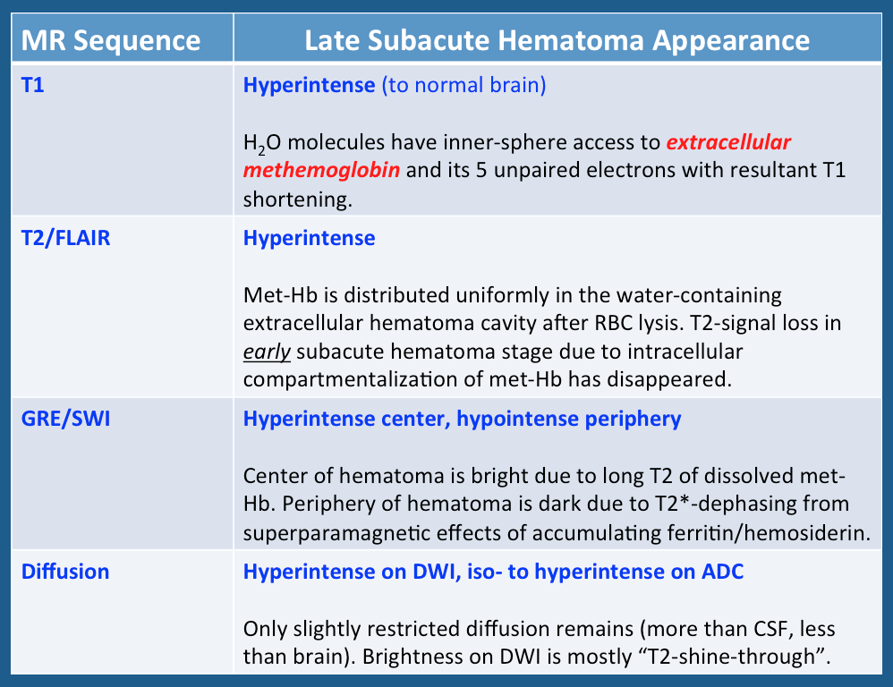
Intracellular vs extracellular methemoglobin mri - Questions and Answers  ​in MRI
Intracellular vs extracellular methemoglobin mri - Questions and Answers ​in MRI

Gradient echo image shows blooming focus. Magnetic resonance imaging... |  Download Scientific Diagram
Gradient echo image shows blooming focus. Magnetic resonance imaging... | Download Scientific Diagram

Neuro-ophthalmology Question of the Week: Magnetic Resonance Imaging —  Neuro-Ophthalmology
Neuro-ophthalmology Question of the Week: Magnetic Resonance Imaging — Neuro-Ophthalmology

Sagittal T2*-weighted gradient-echo image showing a hyperintense area... |  Download Scientific Diagram
Sagittal T2*-weighted gradient-echo image showing a hyperintense area... | Download Scientific Diagram

GRE T2-Weighted MRI: Principles and Clinical Applications

Accuracy of SWI sequences compared to T2*-weighted gradient echo sequences  in the detection of cerebral cavernous malformations in the familial form |  Semantic Scholar
Accuracy of SWI sequences compared to T2*-weighted gradient echo sequences in the detection of cerebral cavernous malformations in the familial form | Semantic Scholar
T2*-weighted imaging - Wikipedia

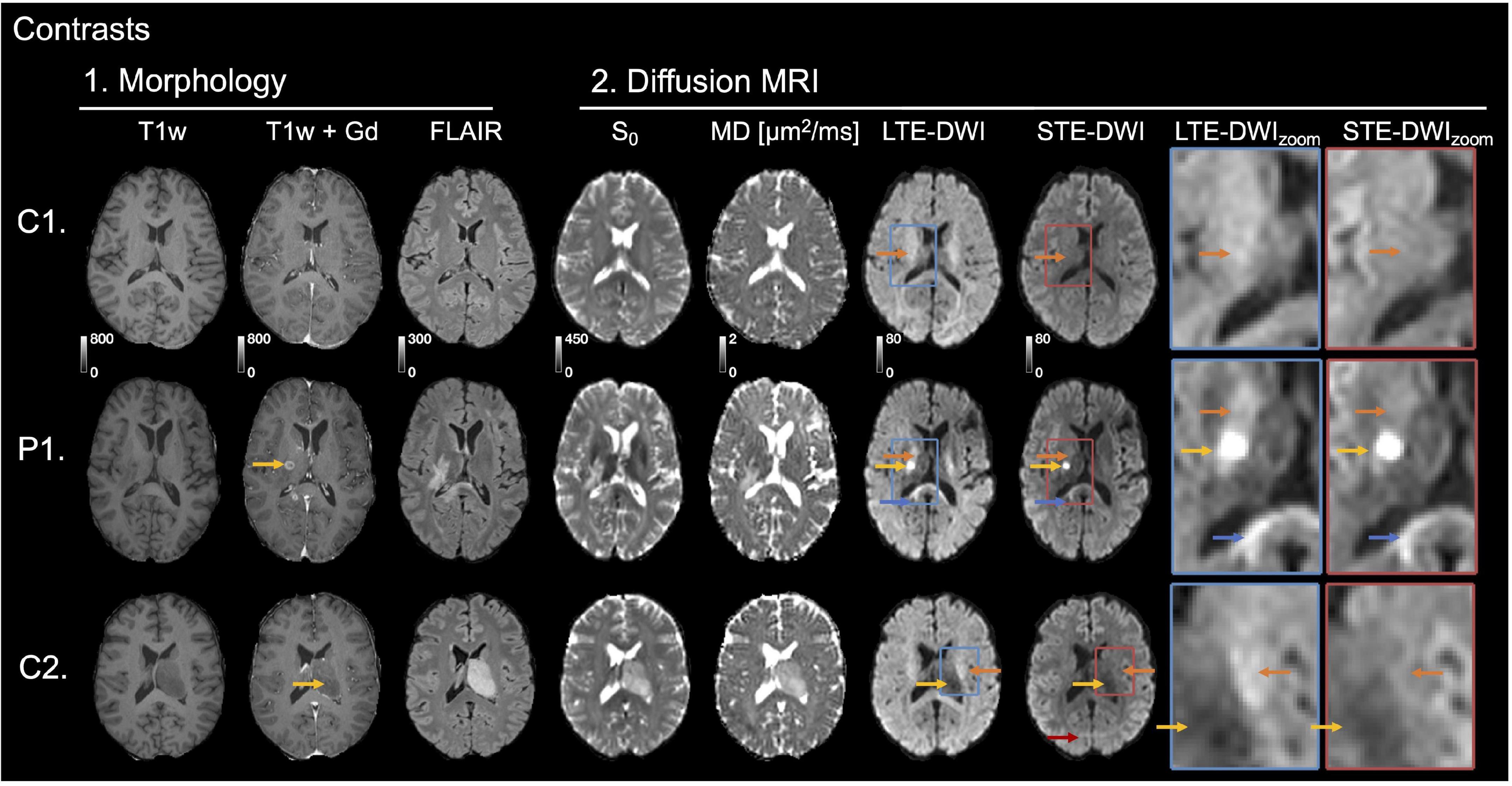
Frontiers | Separating Glioma Hyperintensities From White Matter by  Diffusion-Weighted Imaging With Spherical Tensor Encoding
Frontiers | Separating Glioma Hyperintensities From White Matter by Diffusion-Weighted Imaging With Spherical Tensor Encoding

Fig 2. | Susceptibility-Weighted Imaging for the Evaluation of Patients  with Familial Cerebral Cavernous Malformations: A Comparison with  T2-Weighted Fast Spin-Echo and Gradient-Echo Sequences | American Journal  of Neuroradiology
Fig 2. | Susceptibility-Weighted Imaging for the Evaluation of Patients with Familial Cerebral Cavernous Malformations: A Comparison with T2-Weighted Fast Spin-Echo and Gradient-Echo Sequences | American Journal of Neuroradiology

Hyperintense Thrombus on

EPOS™
EPOS™

EPOS™
EPOS™

EPOS™
EPOS™

GRE T2-Weighted MRI: Principles and Clinical Applications

Gradient Recalled Echo(GRE)

fig 2. | Detection of Intracranial Hemorrhage: Comparison between Gradient- echo Images and b0 Images Obtained from Diffusion-weighted Echo-planar  Sequences | American Journal of Neuroradiology
fig 2. | Detection of Intracranial Hemorrhage: Comparison between Gradient- echo Images and b0 Images Obtained from Diffusion-weighted Echo-planar Sequences | American Journal of Neuroradiology

Flow on GRE sequences - Questions and Answers ​in MRI

MRI Features of Intracerebral Hemorrhage Within 2 Hours From Symptom Onset  | Stroke
MRI Features of Intracerebral Hemorrhage Within 2 Hours From Symptom Onset | Stroke

Hypointensities in the Brain on T2*-Weighted Gradient-Echo Magnetic  Resonance Imaging - ScienceDirect
Hypointensities in the Brain on T2*-Weighted Gradient-Echo Magnetic Resonance Imaging - ScienceDirect